Anatomy
Structures of Your Eye

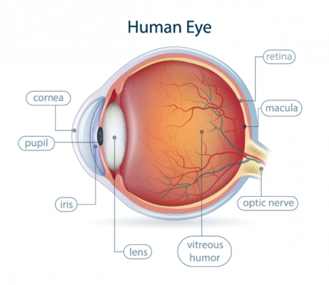
Like a camera, an eye uses light to record images. It has a way to regulate light, a focusing mechanism, and a place where light creates images. However, eyes are much more complex and delicate than even the most expensive and technically advanced camera.
Keeping your eyes in good working order, as with a camera, requires the ability to recognize when they are not working well. That begins with understanding the anatomy of the eye and a few basic terms used in eye care.
Cornea
Light first enters the eye through the cornea, the clear front "window" of the eye. The cornea provides most of the focusing power of the eye.
Pupil
Light travels from the cornea through the pupil, which regulates how much reaches the back of the eye. The pupil is the normally black portion in the center of the eye. It grows larger in low light and smaller when light is more bright and intense.
Iris
The colored portion of the eye is called the iris. It is the muscle that operates the pupil.
Lens
The crystalline lens is a transparent structure located behind the iris that along with the cornea helps to focus light rays entering through the pupil onto the retina.
Retina
Light focuses on a surface called the retina, a saran-wrap thin membrane held to the inside back portion of the eye by a suction-like force. Light receptors called rods and cones are housed within the retina. These nerves translate light into the information sent to the brain along the optic nerve. A special place on the retina, located just above the optic nerve, called the macula, is the most concentrated area of nerves. It is here that most detailed central vision is created.
Optic Nerve
All information from the retina is sent along the optic nerve and into the brain where it is translated into an image.
Rods and Cones
These nerve receptors in the retina are sensitive to different kinds of light. Cone cells provide detail and color or "day" vision and are located primarily in the macula. Rod cells are not color or detail sensitive and provide "night" vision. Rods can be found throughout the retina, but are especially concentrated around its edges to help create the peripheral vision.
Vitreous
The main function of this gel-like fluid is to maintain the shape of the eye. Light coming into the eye through the cornea and lens travels through the vitreous before reaching the retina.
To schedule an appointment, call (509) 457-0107
